Monday, December 1, 2014
Tree of Chordates
Using a combination of anatomical, molecular, and fossil evidence, biologists have developed hypotheses for the evolution for chordate groups. The first transition was the development of a head that consists of a brain at the anterior end of the dorsal nerve cord, eyes and other sensory organs, and a skull. These innovations opened up a completely new way of feeding for chordates: active predation. All chordates with a head are called craniates. The origin of a backbone came next. The vertebrates are distinguished by a more extensive skull and a backbone, or a vertebral column, composed of a serious of bones called vertebrae. These skeletal elements enclose the main parts of the nervous system. The skull forms a case for the brain, and the vertebrae enclose the nerve cord. The vertebrae skeleton is an endoskeleton, made of either flexible cartilage or a combination of hard bone and cartilage. Bone and cartilage are mostly nonliving material. But because there are living cells that secrete the nonliving material, the endoskeleton can grow with the animal. The next major transition was the origin of jaws, which opened up new feeding opportunities. The evolution of lungs or lung derivatives, followed by muscular lobed fins with skeletal support, opened the possibility of life on land. Tetrapods, jawed vertebrates with two pairs of limbs, were the first vertebrates on land. The evolution of amniotes, tetrapods with a terrestrially adapted egg, completed the transition to land. -Biology book page 390
From Ovule to Seed in a Gynosperm
The evolution of vascular tissue solved the terrestrial problems of supporting the plant body and obtaining water and minerals from the soil. However, the challenges of reproduction and dispersing offspring on dry land remained. In contrast to seedless plants, which produce flagellated sperm that need moisture to reach an egg, seed plants--gymnosperms and angiosperms--have pollen grains that carry their sperm producing cells through the air. In addition, the offspring of seedless plants are sent off into the world as haploid, single celled spores that must survive independently as gametophytes before producing the next sporophyte generation. Seed plants launch next generation sporophytes that are ready to grow. In seed plants, a specialized structure within the sporophyte houses all reproductive stages, including spores, eggs, sperm, zygotes, and embryos. In gymnosperms such as pines and other conifers, this structure is called a cone. Cones are modified shoots that serve a reproductive function. -Biology book page 348-349
Ulva
Green algae, which is named for their grass-green chloroplasts, include unicellular and colonial species as well as multicellular seaweeds. Ulva, or sea lettuce, is a multicellular green algae. Like many multicellular algae and all land plants, Ulva has a complex life cycle that includes an alternation of generations. In this type of life cycle, a multicellular diploid form alternates with a multicellular haploid form. Multicellular diploid forms are called sporophytes, because they produce spores. The sporophyte generation alternates with a haploid generation that features a multicellular haploid form called a gametophyte, which produces gametes. In Ulva, the gametophyte and sporophyte organisms are identical in appearance, although they differ in chromosome number. The haploid gametophyte produces gametes by mitosis, and fusion of the gametes begins the sporophyte generation. In turn, cells in the sporophyte undergo meiosis and produce haploid, flagellated spores. The life cycle is completed when a spore settles to the bottom of the ocean and develops into a gametophyte. -Biology book page 336
Bacteria and Archaea
Researchers recently discovered that many prokaryotes once classified as bacteria are actually more closely related to eukaryotes and belong in a domain of their own. As a result, prokaryotes are now classified in two domains: Bacteria and Archaea. Many bacterial and archaeal genomes have now been sequenced. When compared with each other and with the genomes of eukaryotes, these genome sequences strongly support the three-domain view of life. Some genes of archaea are similar to bacterial genes, others are eukaryotic genes, and still others seem to be unique to archaea. Differences between the rRNA sequences provided the first clues of a deep division among prokaryotes. Other differences in the cellular machinery for gene expression include differences in RNA polymerases and in the presence of introns within genes. The cell walls and membranes of bacteria and archaea are also distinctive. Bacterial cell walls contain peptidoglycan, while archaea do not. Furthermore, the lipids forming the backbone of plasma membranes between the two domains. Intriguingly, archaea have at least as much in common with eukaryotes as they do with bacteria. -Biology book page 325
Three Prokaryotes
Some of the diversity of prokaryotes is evident in their external features, including shape, cell walls, and projections such as flagella. These features are useful for identifying prokaryotes as well as helping the organisms survive in their environments, Determining cell shape by microscopic examination is an important step in identifying prokaryotes. Spherical prokaryotic cells are called cocci. Rod-shaped prokaryotes are called bacilli. A third prokaryotic cell shape is spiral, like a corkscrew. Spiral prokaryotes that are relatively short and rigid are called spirilla; those with longer, more flexible cells that cause Lyme disease, are called spirochetes. Nearly all prokaryotes have a cell wall, a feature that enables them to live in a wide range of environments. The cell wall provides physical protection and prevents the cell from bursting in a hypotonic environment. Some prokaryotes have external structures that extend beyond the cell wall. Many bacteria and archaea are equipped with flagella, adaptions that enable them to move about in response to chemical or physical signals in their environment. Biology book page 320-321
Protistan Diversity
Protists are bewilderingly different. Oxygen-using prokaryotes established residence within other, larger cells. These endosymbionts evolved into mitochondria, giving rise to heterotrophic eukaryotes. Autotrophic eukaryotes also arose through endosymbiosis of a prokaryote by a eukaryote after a heterotrophic eukaryote engulfed an autotrophic cynobacterium. If the cynobacterium continued to function within its host cell, its photosynthesis would have provided a steady source of food for the heterotrophic host and thus given it a significant selective advantage. And because the cynobacterium had its own DNA, it could reproduce to make multiple copies of itself within the host cell. In addition, cynobacterium could be passed on when the host reproduced. Over time, the descendants of the origin cynobacterium evolved into chloroplasts. The chloroplast-bearing lineage of eukaryotes later diversified into the autotrophs green algae and red algae. On subsequent occasions during eukaryotic evolution green algae and red algae themselves became endosymbionts following ingestion by different heterotrophic eukaryotes. The heterotrophic host cells enclosed the algal cells in food vacuoles but the algae survived and became cellular organelles. The presence of the endosymbionts, which also had the ability to replicate themselves, gave their hosts a selective advantage. -Biology book page 331
Monday, November 17, 2014
Sea Star Life Cycle
Animals are multicellular, heterotrophic eukaryotes that obtain nutrients by ingestion. Ingestion means eating food. Most animals are diploid and reproduce sexually. Male and female adult animals make haploid gametes by meiosis, and an egg and a sperm fuse, producing a zygote. The zygotes divides by mitosis, forming an early embryonic stage called a blastula, which is usually a hollow ball of cells. In the sea star and most other animals, one side of the blastula folds inward, forming a stage called a gastrula. The internal sac formed by gastrulation becomes the digestive tract, lined by a cell layer called the endoderm. The embryo also has an ectoderm, an outer cell layer that gives rise to the outer covering of the animal and, in some phyla, to the central nervous system. Most animals have a third embryonic layer, known as the mesoderm, which forms the muscles and most internal organs. After the gastrula stage, many animals develop directly into adults. Others, such as the sea star, develop into one or more larval stages first. A larva is an immature individual that looks different from the adult mammal. The larva undergoes a major change of body form, called metamorphosis, in becoming an adult capable of reproducing sexually. Biology book pg 366
Sym Gene Testing
The hypothesized importance of sym genes to plants also predicts that their function should have changed little over time. That is, the sym genes of liverworts should work similarly to those of flowering plants. Scientists tested this prediction by using Agrobacterium to introduce a functional liverwort sym gene into an angiosperm possessing a nonfunctional version of the gene that could not form mycorrhizae. The investigators then applied fungal spores to the roots of transgenic plants and a control group of mutant plants. After several weeks, mycorrhizae were present in the transgenic plants--the result of the liverwort sym gene functioning normally. The control plants had no mycorrhizae. -Biology book pg 360
http://oregonstate.edu/dept/nursery-weeds/weedspeciespage/liverwort/female_sporocarp_750.JPG
http://oregonstate.edu/dept/nursery-weeds/weedspeciespage/liverwort/female_sporocarp_750.JPG
Fungi Life Cycle
Fungal reproduction typically involves the release of vast numbers of haploid spores, which are transported easily over great distances by wind or water. When the hyphae meet, their cytoplasms fuse. But this fusion of cytoplasm is often not followed immediately by the fusion of "parental" nuclei. Thus, many fungi have what is called a heterokaryotic stage, in which cells contain two genetically distinct haploid nuclei. Hours, days, or even centuries may pass before the parental nuclei fuse, forming the usually short-lived diploid phase. Zygotes undergo meiosis producing haploid spores. In asexual reproduction, spore-producing structures arise from haploid mycelia have undergo neither a heterokaryotic stage nor meiosis. Many fungi that reproduce sexually can also produce spores asexually. In addition, asexual reproduction is the only known means of spore production in some fungi, informally known as imperfect fungi. Mold and yeast is also imperfect fungi. The term mold refers to any rapidly growing fungi that reproduces asexually by producing spores, often at the tips of specialized hyphae. The term yeast refers to any single-celled fungus. -Biology book pg 356
Angiosperm Life Cycle
In angiosperms, the sporophyte generation is dominant and produces the gametophyte generation within its body. Meiosis in the anthers of the flower produces haploid spores that undergo mitosis and form the male gametophytes or pollen grains. Meiosis in the ovule produces a haploid spore that undergoes mitosis and forms the few cells of the female gametophyte, one of which becomes an egg. Pollination occurs when a pollen grain, carried by the wind or an animal, lands on the stigma. As in gymnosperms, a tube grows from the pollen grain to the ovule, and a sperm fertilizes the egg, forming a zygote. A seed develops from each ovule. While the seeds develop, the ovary's wall thickens, forming the fruit that encloses the seeds. When conditions are favorable, a seed germinates, which means it began to grow. Biology book pg 351
Plant Life Cycles
Plants have cycles that are very different from ours. Humans are diploid individuals--that is, each of us have two sets of chromosomes, one from each parent. Gametes are the only haploid stage in the human life cycle. Plants have an alternation of generations: the diploid and haploid stages are distinct, multicellular bodies. The haploid generation of a plant produces gametes and is called the gametophyte. The diploid generation produces spores and is called the sporophyte. In a plant's life cycle, these two generations alternate in producing each other. In mosses, as in all nonvascular plants, the gametophytes is the larger, more obvious stage of the life cycle. Ferns, like most plants, have a life cycle dominated by the sporophyte, Today about 95% of all plants, including all seeds plants, have a dominant sporophyte in their life cycle. The life cycles of all plants follow a pattern. -Biology book pg. 346
http://apps.cmsfq.edu.ec/biologyexploringlife/text/chapter19/19images/19-06.gif
http://apps.cmsfq.edu.ec/biologyexploringlife/text/chapter19/19images/19-06.gif
Plant Evolution
After plants originated from an algal ancestor approximately 470 million years ago, early diversification gave rise to seedless, non-vascular plants, including mosses, liverworts, and hornworts. These plants are formally called bryophytes, resemble other plants in having apical meristems and embryos that are retained on the parent plant, but they lack true roots and leaves. Without lignified cell walls, bryophytes with an upright growth habit lack support. The origin of vascular plants occurred about 425 million years ago. Their lignin-hardened vascular tissues provide strong support, enabling stems to stand upright and grow tall on land. Two clades of vascular plants are informally called seedless vascular plants: lycophytes and widespread monilophytes. The first vascular plants with seeds evolved about 360 million years ago. Seeds and pollen are key adaptions that improved the ability of plants to diversify in terrestrial habitats. A seed consists of an embryo packaged with a food supply within a protective covering. -Biology book pg. 344
http://antranik.org/wp-content/uploads/2011/06/plant-evolution.jpg
http://antranik.org/wp-content/uploads/2011/06/plant-evolution.jpg
Monday, October 20, 2014
DNA Profiling
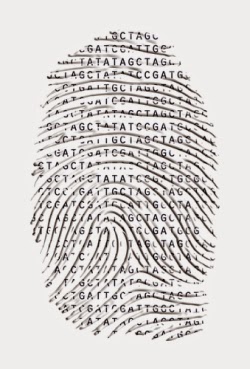
Modern DNA technology methods have rapidly transformed the field of forensics. Forensics is the scientific analysis of evidence for crime scene investigations and other legal proceedings. The most important application of forensics is DNA profiling. DNA profiling is the analysis of DNA samples to determine whether they came from the same individual. First, DNA samples are isolated from the crime scene, suspects, victims, or other evidence. Second, selected markers from each DNA sample are copied many times, producing a large sample of DNA fragments. And last, the amplified DNA markers are compared, providing data about which samples are from the same individual. -Biology book pg. 242
Genetically Modified Organisms
For many years, scientists have selectively bred agricultural crops to make them more useful. In recent years, DNA technology has quickly replaced traditional breeding programs. Genetic engineers have produced many different varieties of genetically modified organisms. GMO's are organisms that have acquired one or more genes by artificial means. A common vector used to introduce new genes into plant cells is a plasmid from the soil bacterium Agrobacterium tumefaciens called the Ti plasmid. With the help of a restriction enzyme and a DNA ligase, the gene for the desired trait is inserted into a modified version of the plasmid. Then the recombinant plasmid is put into a plant cell, where the DNA carrying the new gene integrates into one of the plant's chromosomes. Finally, the recombinant cell is cultured and grown into a plant. With an estimated one billion people facing malnutrition, GMO crops may be able to help many hungry people by improving food production, shelf life, pest resistance, and the nutritional value of crops. -Biology book pg. 239
http://s36.photobucket.com/user/seahawks940/media/plasmid.jpg.html
Sunday, October 19, 2014
Gene Cloning
To start off the process of gene cloning, biologists isolate two kinds of DNA, a bacterial plasmid and the DNA from another organism that includes the gene that codes for protein V. The DNA containing gene V could come from different bacterium, a plant, a nonhuman animal, or even human tissue cells. Both the plasmid and the gene V source DNA are treated with an enzyme that cuts. The cells DNA is cut with the same enzyme. The cut DNA from both sources are mixed. DNA ligase is added joining the two DNA molecules by way of covalent bonds. The recombinant plasmid is taken up by a bacterium through transformation. The recombinant bacterium then reproduces to form a clone of cells, each carrying a copy of gene V. This step is actual gene cloning. This process can be used to create a number of products. Copies of gene itself can be the immediate product, used in plants. Other times, the protein product of the cloned gene is harvested and used, to make "stoned-washed" blue jeans. A protein can also be used to dissolve blood clots in heart attack therapy.
Colon Cancer
In just one year alone, more than 100,000 Americans will get colon cancer. The colon is the main part of the large intestine. It takes more than one somatic mutation to produce a full-fledged cancer cell. Colon cancer begins when an oncogene arises or is activated through mutation, causing unusually frequent division of apparently normal cells in the colon lining. Later, additional DNA mutations cause the growth of a small benign tumor in the colon wall. Still more mutations eventually lead to formation of a spreading tumor. The requirement for several mutations explains why cancers can take a long time to develop. The actual number of mutations is usually around six. Multiple changes must occur at the DNA level for a cell to become fully cancerous. Once a cancer promoting mutation occurs, it is passed to all the descendants of the cell carrying it. The fact that more than one somatic mutation is generally needed to produce a full-fledged cancer cell may help explain why the incidence of cancer increases with age.
http://www.zo.utexas.edu/faculty/sjasper/images/19.15.gif
Thursday, October 16, 2014
Rosalind Franklin
Rosalind Franklin was born in London England in 1920. As a little girl she was astoundingly smart for her age. Her parents sent her to St. Paul's Girls School where she graduated a year early and went to Cambridge University to major in physics and chemistry in 1938. There, she was introduced to x-ray crystallography. She then joined the war effort doing research on coal. Her research led to better gas masks, publishing five landmark papers, and being awarded her PhD. After the war she landed a research position in Paris and took on x-ray diffraction. She spoke in conferences and published articles in journals. Over a short period of time, she exceeded unsafe levels of x-ray radiation and was put out of the lab for a few months. So she decided to attend King College London where she was hired by JT Randall to discover DNA in January 1951. When she went in she was supposed to be Maurice Wilkins' assistant but he was away on vacation. He come back to find that Franklin took over his lab as well as his PhD student, Raymond Gosling, which was all due to miscommunication from the leader Randall.
James Watson comes to London to study DNA where he teams up with Francis Crick to make a model of the DNA structure. Meanwhile, Franklin discovered two distinct forms of DNA, A and B. In November 1951, Crick sent Watson to Franklin's lecture about her A and B discovery to try and get information to help them build they're DNA model. Within a week after her lecture, they invite her to see they're model at Cambridge University where they were embarrassed after she told them it was completely wrong. So Lawrence Bragg, the head of the Cavendish Laboratory, told them they couldn't make anymore models because they humiliated him.
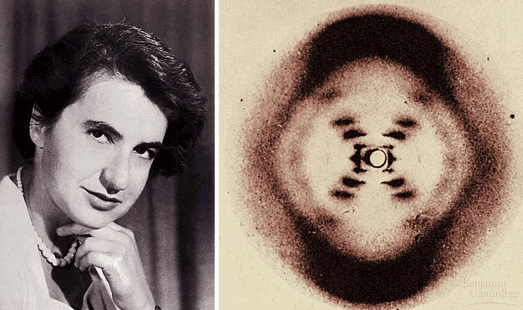
In May 1952, Franklin sets up an x-ray diffractometer to take a better picture of the B form. In her results, she gets a clearer, sharper image of B and labels it Photo 51 and puts it safely away while she works on form A. At the end of the year, she decides to leave Cambridge University due to the rude people but kept up her work and study. Some how in the mix of her leave, her Photo 51 gets leaked to Wilkins. Linus and his son Peter Pauling did model building and knew almost as much as Franklin did. Watson warned Franklin that Pauling was going to beat them to the DNA secret if she didn't go in with him and Crick and publish it but she said no. Wilkins shows Watson Photo 51 and Watson shows Crick and they begin making another model with permission from Bragg on February 4th, 1953. On February 28th, 1953, the model was complete. Franklin was amazed and didn't understand how they knew without her detailed photos. Articles were released in Nature on April 25th, 1953 stating Crick and Watson's untruthful discovery and gave no mention of Franklin's photo.
After leaving Cambridge, Franklin attended Birkbeck University of London where she led the virus research lab from 1953-1958. There, she collaborated with Aaron Klug working out the complex structure of a virus and locating it's infectious element. Together, they won a Nobel Prize. In 1956, she celebrated her 36th birthday by visiting universities in California and climbing Mt. Whitney. At the end of her trip, she began to suffer from severe abdominal pains. When she returned home, she was diagnosed with cancer believed to be caused from years of x-ray radiation. Even with the disease, she still studied every day, feeling as if she were too busy to die. She soon died on April 16th, 1958.
In 1962, James Watson, Francis Crick, and Maurice Wilkins won the Nobel Prize for the discovery of the structure of DNA. None of the three acknowledged Rosalind Franklin for her unknowing contribution to the discovery. In 1968, James Watson published a best seller book entitled The Double Helix. In the book, he admits he wouldn't have ben able to finish his work without her findings and that he used them without permission. When Aaron Krug was awarded the Nobel Prize, he honored her contribution to the study of DNA in mentioning her name and her hard work.
Tuesday, October 14, 2014
Charles Darwin
Charles Darwin introduced the theory of evolution to the world more than 200 years ago. He published a book entitled On the Origin of Species by Means of Natural Selection. In 1841 Charles Darwin set out on a ship called the Beagle to explore the South American coast. On his voyage he took detailed notes and collected fossils, bones, plants, insects, and many other things. From his studies, he came to realize that the world isn't just 6,000 years old, as the book of Genesis states in the bible, but that it is over hundreds of thousands of years old. When he returns home, he put all of his evidence that he collected over his 5 year trip and seen that he discovered 69 different species of beetles as well as different species of butterflies, dragon flies, birds, and many other organisms including plants. He gave a skull head he found to Richard Owen he informed him that his finding was the skull of a ground sloth that is a species that is now extinct that lived over 10,000 years ago.
Sunday, September 21, 2014
Cancer
What is cancer? Cancer is a disease characterized by the presence of malignant tumors in the body. Malignant tumors can spread into surrounding tissues and invade other parts of the body displacing normal tissue and interrupting organ function as it grows. A tumor grows from a single cancer cell. It starts when the cell undergoes transformation, converting a normal cell to a cancer cell. A transformed cell grows abnormally and the immune system usually recognizes it and destroys it. If the cell somehow avoids destruction, it may multiply to form a tumor. A tumor is a mass of abnormally growing cells with otherwise normal tissue. If the abnormal cells remain in their original spot, then a benign tumor has formed. These tumors can be dangerous if they spread to the brain but if they maintain size and location they can be left alone. If needed, they can be completely surgically removed. What you don't want is for the cancer cells to spread through lymph and blood vessels to other parts of the body. This process is called metastasis. Unfortunately, cancer cells are not density-dependent. If they were, they would stop multiplying when they have filled a single layer. Instead they keep dividing, forming a clump of overlapping cells.
http://2.bp.blogspot.com/-bLMsNN3xWmA/UDV2oRDnB3I/AAAAAAAAJDY/vTXEmUk0mKA/s640/The+growth+of+breast+cancer.jpg
http://2.bp.blogspot.com/-bLMsNN3xWmA/UDV2oRDnB3I/AAAAAAAAJDY/vTXEmUk0mKA/s640/The+growth+of+breast+cancer.jpg
Down Syndrome
An extra copy of chromosome 21 is what causes down syndrome. In a normal human complement, there are 23 pairs of chromosomes. But what is down syndrome and what causes this to happen? Down syndrome is a genetic condition in which a person has 47 chromosomes instead of 46, which is the normal amount. This is also a related to trisomy 21. Trisomy 21 is when there are three number 21 chromosomes, making 47 chromosomes in total. It is the most common chromosome number abnormality and the most common serious birth defect in the U.S. It affects about one out of every seven hundred children. Some of the characteristics of a person with down syndrome possess a round face, a skin fold at the inner corner of the eye, a flattened nose bridge, and small, irregular teeth. They're also short and have heart defects. As well as susceptibility to respiratory infections, leukemia, and Alzheimer's disease. Generally, people with down syndrome have a shorter life span than others. The older a woman is when she has a baby, the higher the percentage is for her to have a child with this chromosome defect. Less than .05% in women under the age of 30, 1% for mothers at age 40, and the percentage is even higher for older mothers.
http://anthro.palomar.edu/abnormal/images/Down_Syndrome_Karyotype.jpg
http://anthro.palomar.edu/abnormal/images/Down_Syndrome_Karyotype.jpg
Thursday, September 18, 2014
Chemical Cycling and Energy Flow in an Ecosystem
Did you know that we as humans inhale oxygen and exhale carbon dioxide? And did you know that plants and most other organisms do the exact same thing? But how? The ecosystem works in an amazing way. The sun is the light energy for the entire ecosystem. It keeps the plants growing, which are the producers for the ecosystem. Trees absorb water and minerals through its roots from the soil and its leaves take in carbon dioxide from the air. The leaves then release oxygen into the air. The consumers of the ecosystem are the animals. They eat the plants and other animals. Decomposers such as, worms, fungi, and bacteria return chemicals back to the soil by changing complex matter into simpler chemicals. The most basic chemicals necessary for life are carbon dioxide, oxygen, water, and various minerals. As you can see, the ecosystem is a big, never ending cycle of producers and consumers. Nothing can do anything without the step before it and after it.
Saturday, September 13, 2014
Alcoholic Fermentation
Alcoholic fermentation is one of two pathways, lactic fermentation being the other. Both fermentations occur in two stages. In the first stage, yeast breaks down glucose into pyruvate through a process called glycolysis. Glycolysis is the first stage in cellular respiration in all organisms. It is a process that cells use to break down glucose for energy. Pyruvate is what happens as a result of glycolysis. A carbon dioxide molecule is removed from the pyruvic acid leaving a two-carbon compound. In the second stage, two hydrogen atoms from NADH and N+ are combined to the two-carbon compound to create ethyl alcohol. The NADH is oxidized to make NAD+. Oxidation is when a substance loses electrons. NADH is the reduced state of NAD+.
- In Biology 101, we took 1 tablespoon of sugar and a small packet of yeast and mixed them together in warm water. Next we put it inside a 20oz plastic bottle and tightly covered the top of the bottle with a rubber glove and let it sit for about 20 minutes. The glove started to expand from the carbon dioxide being released. This process is used to create wine, beer, and bread. This example is a representation of alcoholic fermentation.
Biology
Biology is an interesting subject. It's one of its own in many ways. It tells you how the whole world; from the air we breathe to the germs on our counters; is made up of molecules and cells that you can't see with the naked eye. Many scientists have dedicated their lives to discovering things and how they work. In November of 1859, Charles Darwin wrote a book called On The Origin of Species by Means of Natural Selection which was about evolution. Evolution is the process that explains how life on Earth was billions of years ago in relation to now. Peter Agre was another scientist. He made the discovery of aquaporins. This discovery lead him and his partner, Roderick MacKinnon, to get the Nobel Prize in Chemistry in 2003. An aquaporin is a transport protein in the cell membrane that allows water to pass through to the membrane. These scientists, and every other scientist, has to be very open minded about things and expect anything to happen.


http://upload.wikimedia.org/wikipedia/commons/2/2e/Charles_Darwin_seated_crop.jpg
http://upload.wikimedia.org/wikipedia/commons/8/8d/Peter_Agre.jpg
http://nihrecord.nih.gov/newsletters/2004/10_12_2004/images/STETLECT.jpg


http://upload.wikimedia.org/wikipedia/commons/2/2e/Charles_Darwin_seated_crop.jpg
http://upload.wikimedia.org/wikipedia/commons/8/8d/Peter_Agre.jpg
http://nihrecord.nih.gov/newsletters/2004/10_12_2004/images/STETLECT.jpg
Sunday, September 7, 2014
Eukaryotic Cells
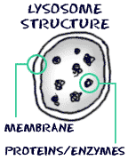
Between prokaryotic cells and eukaryotic cells, eukaryotic cells are my favorite. They're more interesting than prokaryotic cells. They contain more parts to their cells and are larger. Lysosomes and centrosomes are the only organelles that are found in eukaryotic cells that are not found in prokaryotic cells. Lysosomes are recyclers for the cells. They also work to make cells renew themselves. They are made by rough endoplasmic reticulum and work through the Golgi apparatus. Protists take in food particles into food vacuoles which digests the food. Lysosomes connect with food vacuoles and digest the food. Then nutrients are released into the cytosol. Our white blood cells take in bacteria and then get rid of them using lysosomes. A lysosome connects with enclosed damaged organelles or small amounts of cytosol located in membrane sacs and destroys its contents. As a result, it makes organic molecules available for reuse. Diseases such as Tay-Sachs disease and Gaucher disease are two examples from lysosome storage deficiencies.


Subscribe to:
Comments (Atom)


